Back Muscles Anatomy / Diagram Back Muscles Human Anatomy Diagram Shoulder Muscle Anatomy Muscle Diagram Shoulder Anatomy : These are the muscles that are farther from the surface, closer to the internal organs and the spine.
Back Muscles Anatomy / Diagram Back Muscles Human Anatomy Diagram Shoulder Muscle Anatomy Muscle Diagram Shoulder Anatomy : These are the muscles that are farther from the surface, closer to the internal organs and the spine.. As a general group, they extend from the neck to the sacrum and fulfill a basic and fundamental function: The anatomy of the back refers to the muscles of the back, as well as the bones of the scapulae, ribcage, and spine.covering an expanse from the neck to the tailbone, the back muscles are responsible for a broad range of functions, from extending the spine to shrugging the shoulders.these muscles facilitate movement by attaching to one or more bones of the back, either to the spinous processes. Lumbar spine anatomy video understanding the anatomy of your lower spine can help you communicate more effectively with the medical professionals who treat your lower back pain. These structures work together to support the body, enable a range of movements, and send messages from the brain to the. All these muscles are therefore associated with movements of the upper limb.
The muscles of the lower back help stabilize, rotate, flex, and extend the spinal column, which is a bony tower of 24 vertebrae that gives the body structure and houses the spinal cord.the spinal. This blog post article is an overview of the muscles of the lumbar spine of the trunk. These are the muscles that are farther from the surface, closer to the internal organs and the spine. Three types of back muscles that help the spine function are extensors, flexors and obliques. The extrinsic back muscles are located in the back, but act to produce movements of the shoulder and assist respiration.
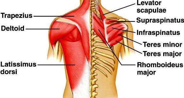
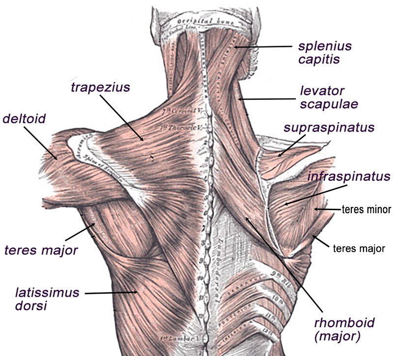
Back Muscles Science Online from www.online-sciences.com The small muscles of the vertebrae (the multifidi and rotators) help rotate, extend, and side bend the back. For more anatomy content please follow us and visit our website: The muscles of the chest and upper back occupy the thoracic region of the body inferior to the neck and superior to the abdominal region and include the muscles of the shoulders. They provide movements of the spine , stability to the trunk, as well as the coordination between the movements of the limbs and trunk. This is a tutorial to quickly s. Also in the back are the teres major, teres minor, trapezius, rhomboids, erector spinae Lower back muscle anatomy includes the multifidus, longissimus, spinalis, and quadratus lumborum. Several small muscles in the cervical area of the vertebral column are also important.
The muscles of the chest and upper back occupy the thoracic region of the body inferior to the neck and superior to the abdominal region and include the muscles of the shoulders.
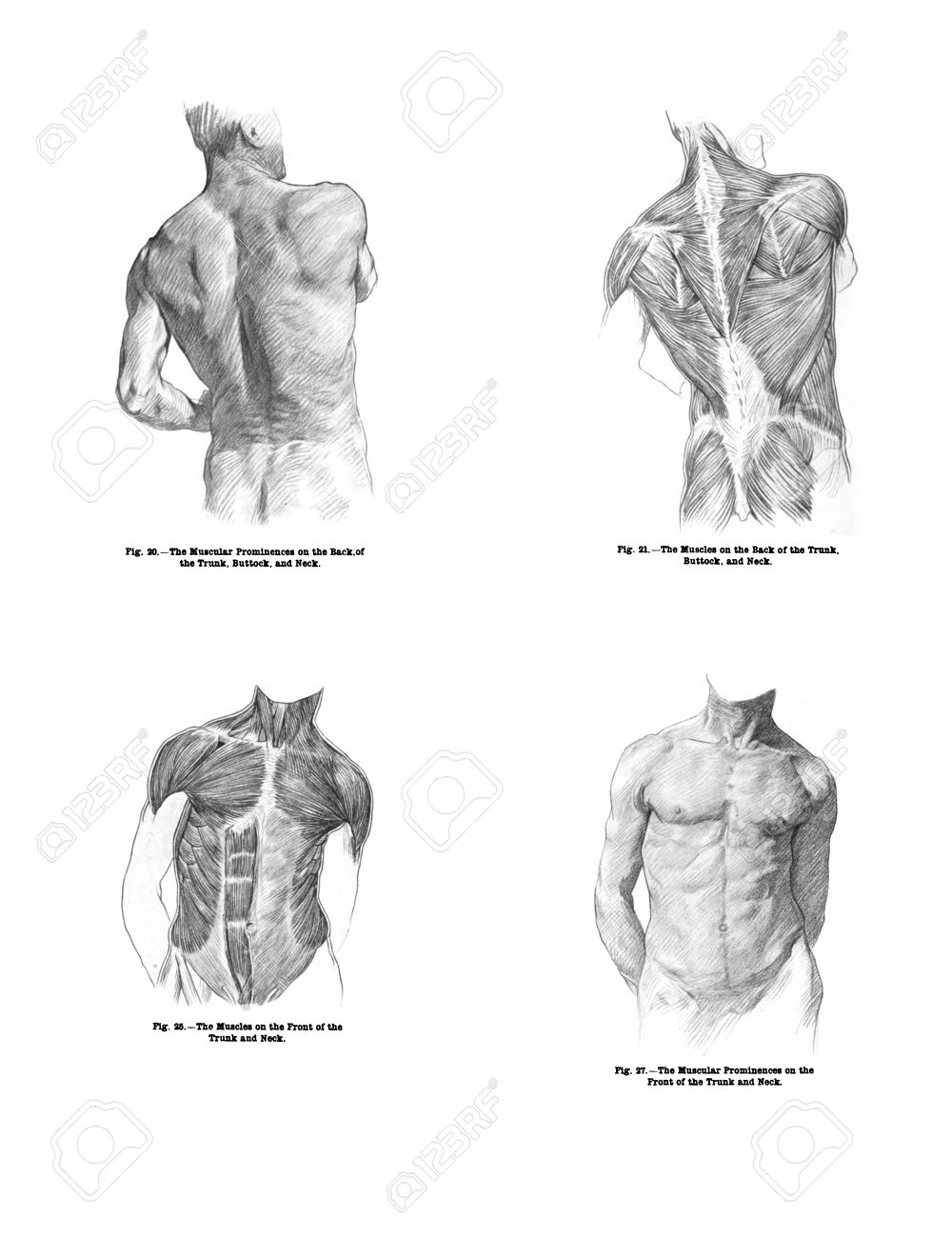
The upper back has the most structural support, with the ribs attached firmly to each level of the thoracic spine and very limited movement. Body muscle parts 12 photos of the body muscle parts body parts and muscle names, human body muscle parts, muscle parts in your body, upper body muscle parts, v rod muscle body parts, human muscles, body parts and muscle names, human body muscle parts, muscle parts in your body, upper body muscle parts, v rod … Anatomy of the back muscles. Also in the back are the teres major, teres minor, trapezius, rhomboids, erector spinae The extrinsic back muscles are located in the back, but act to produce movements of the shoulder and assist respiration. The intricate anatomy of the back provides support for the head and trunk of the body, strength in the trunk of the body, as well as a great deal of flexibility and movement. Intermediate and deep muscles of the back. Three types of back muscles that help the spine function are extensors, flexors and obliques. The muscles of the chest and upper back occupy the thoracic region of the body inferior to the neck and superior to the abdominal region and include the muscles of the shoulders. These muscles give height and breadth to back development. The extrinsic muscles that are associated with upper extremity and shoulder movement, and the intrinsic muscles that deal with movements of the vertebral column. The anatomy of the back refers to the muscles of the back, as well as the bones of the scapulae, ribcage, and spine.covering an expanse from the neck to the tailbone, the back muscles are responsible for a broad range of functions, from extending the spine to shrugging the shoulders.these muscles facilitate movement by attaching to one or more bones of the back, either to the spinous processes. Several small muscles in the cervical area of the vertebral column are also important.
(2017, elsevier) should be consulted. Muscle anatomy basics 12 photos of the muscle anatomy basics basics of muscle anatomy, muscle anatomy basics, human muscles, basics of muscle anatomy, muscle anatomy basics. The extrinsic back muscles are located in the back, but act to produce movements of the shoulder and assist respiration. The back muscles are divided into two large groups: The upper back has the most structural support, with the ribs attached firmly to each level of the thoracic spine and very limited movement.
Muscles Of The Back from www.biologycorner.com The surface muscles of the upper back include the trapezius muscles (traps) and posterior deltoids. Extrinsic muscles of the back. Learn all about the muscles of the back in this 3d video anatomy tutorial. Back, musculoskeletal, tutorials by admin december 6, 2011 5 comments. The lumbar region of the spine, more commonly known as the lower back, is situated between the thoracic, or chest, region of the spine, and the sacrum. The back consists of the spine, spinal cord, muscles, ligaments, and nerves. The back muscles are anatomically layered into superficial (extrinsic) and deep (intrinsic) muscles. We hope this picture anatomy of back muscles diagram can help you study and research.
Learn all about the muscles of the back in this 3d video anatomy tutorial.
Back muscles the muscles of the back are a group of strong, paired muscles that lie on the posterior aspect of the trunk. Also in the back are the teres major, teres minor, trapezius, rhomboids, erector spinae They provide movements of the spine , stability to the trunk, as well as the coordination between the movements of the limbs and trunk. Intermediate and deep muscles of the back. To control the posture of the entire body. Related posts of diagram of female back muscles body muscle parts. The erector spinae muscles, for example, extend the back (bend it backward) and side bend the back. The small muscles of the vertebrae (the multifidi and rotators) help rotate, extend, and side bend the back. We think this is the most useful anatomy picture that you need. The back anatomy includes the latissimus dorsi, trapezius, erector spinae, rhomboid, and the teres significant. Intermediate and deep muscles of the back. Since the all the back muscles originate in embryo (fetus) form by locations other than the back, muscles in the superficial, as well as, intermediate groups, are extrinsic muscles. The lumbar region of the spine, more commonly known as the lower back, is situated between the thoracic, or chest, region of the spine, and the sacrum.
Back muscles are divided into two specific groups: To control the posture of the entire body. The muscles of the back can be arranged into 3 categories based on their location: Lower back muscle anatomy includes the multifidus, longissimus, spinalis, and quadratus lumborum. Anatomynote.com found anatomy of back muscles diagram from plenty of anatomical pictures on the internet.
4 Views Of The Human Back Muscles And Torso From Out Of Print Stock Photo Picture And Royalty Free Image Image 11309096 from previews.123rf.com The intrinsic back muscles are found deeper to the extrinsic muscles, separated from them by the thoracolumbar fascia. The deep muscles develop in the back called intrinsic muscles. Related posts of diagram of female back muscles body muscle parts. The muscles of the back can be arranged into 3 categories based on their location: As a general group, they extend from the neck to the sacrum and fulfill a basic and fundamental function: Related posts of back muscles chart muscle anatomy basics. Intermediate and deep muscles of the back. On this page, youll learn about each of these muscles, their locations, and functional anatomy.
This is a tutorial to quickly s.
Intermediate and deep muscles of the back. Several small muscles in the cervical area of the vertebral column are also important. Intermediate and deep muscles of the back. Learn all about the muscles of the back in this 3d video anatomy tutorial. Anatomynote.com found anatomy of back muscles diagram from plenty of anatomical pictures on the internet. 3d video anatomy tutorial on the intermediate and deep muscles of the back. (2017, elsevier) should be consulted. Also in the back are the teres major, teres minor, trapezius, rhomboids, erector spinae The lumbar region of the spine, more commonly known as the lower back, is situated between the thoracic, or chest, region of the spine, and the sacrum. Since the all the back muscles originate in embryo (fetus) form by locations other than the back, muscles in the superficial, as well as, intermediate groups, are extrinsic muscles. All these muscles are therefore associated with movements of the upper limb. Back muscles are divided into two specific groups: Related posts of back muscles chart muscle anatomy basics.